Medical Retina Services and Surgery

The retina is the thin piece of tissue lining the innermost wall of the eye. It turns light rays entering our eyes into the electrical signals that become the images we “see”.

Some medical conditions – and even ageing – can negatively affect the retina’s function. These include issues like age-related macular degeneration or AMD retina, which can lead to vision loss. 

At ERS Suntec City, we can offer retinal disease treatment and consultation for a variety of retina damage symptoms.
Dry AMD occurs when the macula thins overtime due to wear and tear. It is the more common form of AMD, with around 90% of patients having it. While it rarely causes severe vision loss, it can still lead to wet AMD, which is more severe.

Symptoms of dry AMD

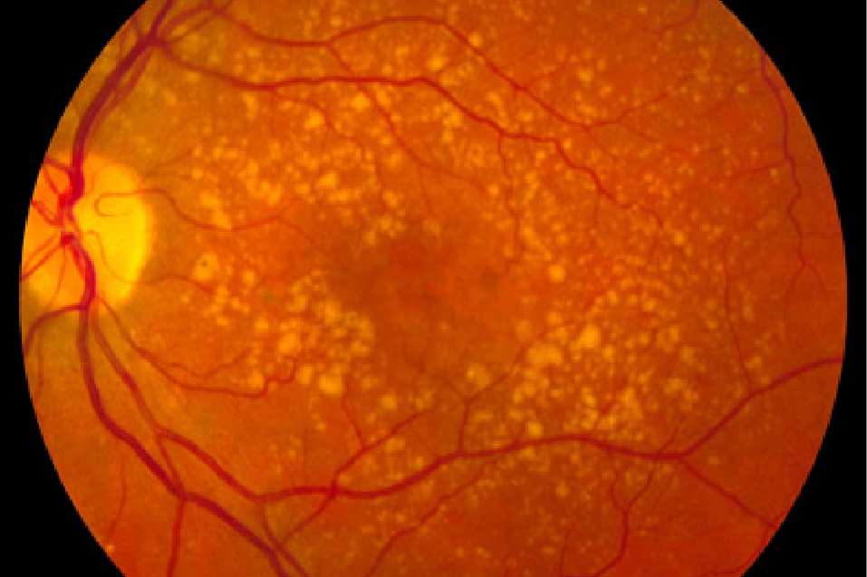
Dry AMD can be asymptomatic in its early stages, but it can also eventually show in a dark spot in your central vision. Even when this spot is very small and light, it may be wise to address the situation quickly to avoid it taking more of your central vision.

Causes of dry AMD

Dry AMD can be caused by either normal ageing or environmental factors. Generally, it happens as waste deposits called drusen form under the macula and cause it to thin.

Treatment options for dry AMD

The options for dry age related macular degeneration treatment range from vitamin therapy to lifestyle changes. Trials are also ongoing to find drugs that can slow or stop the breakdown of the macula. An example is brimonidine, often administered as eye drops.
Wet AMD is the rarer, more dangerous form of macular degeneration. It happens to about 10% of patients and can lead to permanent vision loss. The name comes from abnormal blood vessels leaking blood or other fluids under the retina. These blood vessels may break through your retinal layers and appear as spots in your vision if portions of the retina are damaged. 

Symptoms of wet AMD

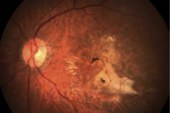
Wet AMD symptoms are generally related to abnormal or distorted central vision. For instance, when looking at a grid of straight lines, you may find the central line in your view wavy. Blurry or dark central vision can also be a symptom of wet AMD.

These symptoms can worsen over time and can lead to irreversible vision loss if untreated. This is why it is generally advised to get screened for and potentially treat wet AMD once such signs appear.

Causes of wet AMD

Wet AMD is often caused by the growth of abnormal blood vessels under the retina. These vessels are often very delicate and break easily, leaking blood and fluids under the retina. The fluid builds up and causes the retina to bulge away and detach from the back of the eye.

Treatment options for wet AMD

There are two main treatment options for wet AMD: anti-VEGF injections and photodynamic therapy (PDT) with verteporfin (Visudyne). 

Anti-VEGF injections

Anti-VEGF (vascular endothelial growth factor) injections work by discouraging the growth of the abnormal blood vessels. The medication that does this is injected directly into the eye. Possible medications used for this are Lucentis, Eylea, and Beovu.

Upcoming anti-VEGF drugs

There are also new anti-VEGF drugs being tested. Abicipar and RGX-314 are just two examples, with the latter even using gene therapy. Refillable and implantable port-delivery methods are also being considered as an alternative to eye injections.

 Photodynamic therapy with verteporfin

Photodynamic therapy (PDT) with verteporfin (Visudyne) is now being used in combination with anti-VEGF treatments. Visudyne is injected into the blood through a vein in the arm, which travels to the back of the eye. There, it’s activated with a cold laser, which causes it to close the leaky blood vessels that are the source of wet AMD.

Visudyne is a multi-course therapy given about every 3 months, for as many times as it is required to address the abnormal blood vessels.
Central serous chorioretinopathy or CSC is a disorder where fluid builds up under the retina. When this happens, a serous or fluid-filled bulge pushes the retina away from the back of the eye. This detachment of the retina can lead to vision loss.

Symptoms of CSC

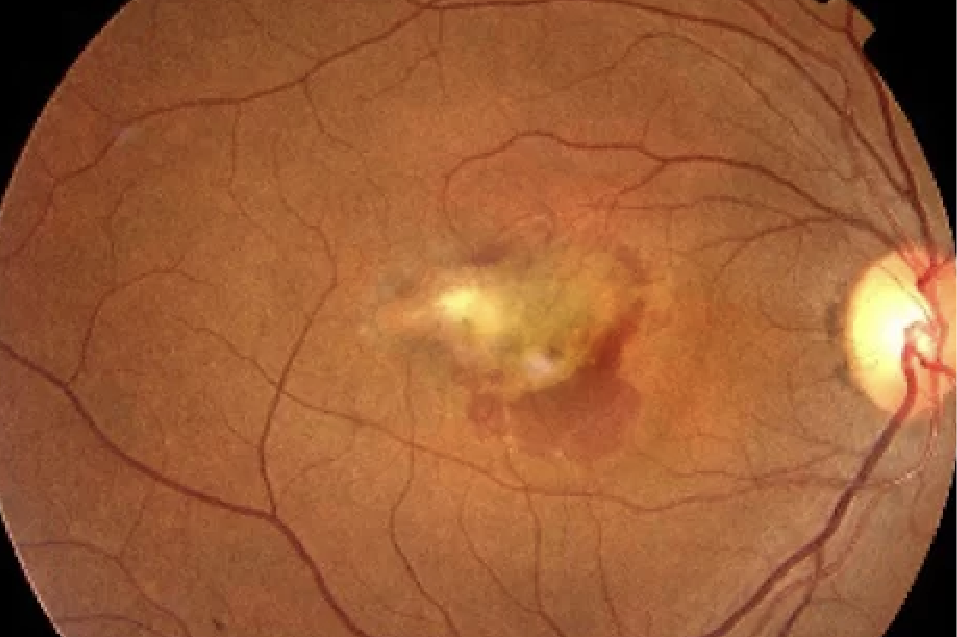
Central serous chorioretinopathy symptoms may include blurry central vision, distortion of straight lines, and micropsia, where an object looks smaller or more distant to an affected eye than to a normal one. However, CSC can also be asymptomatic if the affected areas lie outside of the macula.

Causes of CSC

The exact cause of CSC is still unknown, but it has been associated with the use of corticosteroids found in nose sprays and topical anti-inflammatory creams. Some also suggest that people suffering from emotional stress or prone to producing stress hormones are more likely to get the condition.

High blood pressure, heart disease, and exposure to certain drugs may also make you more likely to develop CSC. Rare congenital anomalies such as focal choroidal excavation and optic disc pit may be linked to a predisposition to CSC as well.

Types of CSC

The two types of CSC are the acute and chronic. Because acute CSC often resolves within a few months without treatment, only chronic CSC is usually treated. People with chronic CSC are those who still suffer its symptoms after 3 months following onset.

Treatment options for CSC

The treatments for CSC range from laser to injected or oral medications such as Eplerenone.

Cold laser, also called photodynamic therapy (PDT), can also be used with the drug verteporfin to close the source of the leakage in the eye. In PDT, verteporfin (Visudyne) is injected into the blood through a vein in the arm, which travels to the back of the eye. There, it’s activated with a cold laser to address the source of the leakage in chronic CSC and prevent future recurrences.

As for oral medications, Eplerenone may be used – although side effects can limit its application.
PCV is a disease where abnormal blood vessels appear in the choroid, a part of the eye full of connective tissues. When left untreated, PCV can damage the retina nearest that area. PCV vessels tend to look like orange bulges, also called bunches of grapes or pearl strings.

Symptoms of PCV

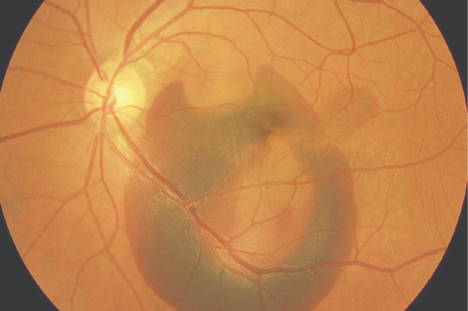
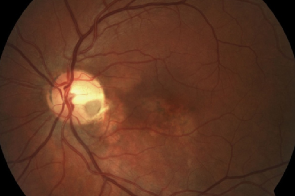
The symptoms of PCV include blurry vision or partial blind spots, usually near the centre of your vision. Scotoma or an abnormally distorted part of otherwise clear vision is another common symptom. Severe cases of PCV may result irreversible scarring and loss of central vision if left untreated.

Causes and risk factors of PCV

The exact cause of PCV is still unknown, but some suggest that risk factors for it include age, genetics, and ethnicity (because it occurs more often in Asian and African patients). Those over 60 years old also appear more likely to get the condition.

Tests and diagnosis for PCV

PCV can be diagnosed by an experienced ophthalmologist through tests such as a dilated eye examination, fluorescein and indocyanine green (ICG) angiography, as well as optical coherence tomography (OCT).

The best retina specialist for such cases will typically perform these tests along with an evaluation of your medical history and your current condition to diagnose PCV.

Treatment options for PCV

The treatment options for PCV are similar to the ones for wet AMD. They include intravitreal injections of anti-VEGF to prevent abnormal blood vessel growth as well as photodynamic therapy (PDT) to seal leaky vessels. Cases with large haemorrhages in the eye may also require a surgical vitrectomy or removal of the eye’s clear vitreous gel.
RVO is a disease where one of the four retinal veins is blocked by a blood clot, preventing blood from draining away from the retina as it should. A retinal vein occlusion is thus a blockage in a blood vessel, interrupting the normal supply of oxygen and nutrients to the eye.

While it is sometimes referred to as an “eye stroke” due to the vision loss it can cause, RVO is not classified as a stroke.

Symptoms of RVO

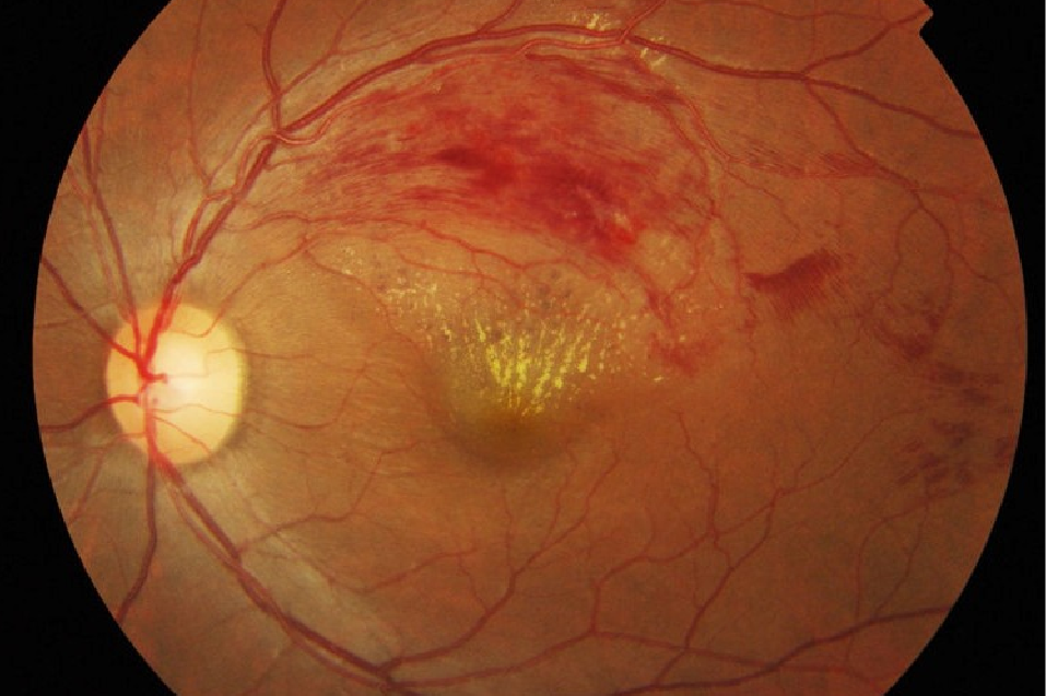
The symptoms of RVO depend on the severity of the blockage of the affected retinal vein. Milder cases may be asymptomatic, while partial or serious vision loss may occur in more severe cases of RVO.

Here are some of the most common symptoms of RVO: 

  • Sudden or gradual vision loss
  • A blind spot for the affected eye
  • Increased pressure or discomfort in the eye
  • Reddishness in the eyes
  • Floaters

Causes of RVO

Retinal vein occlusion can be caused by blood clots getting stuck in the narrow veins of the eye. This means that people with high blood pressure, diabetes, high cholesterol levels, and smoking habits are at higher risk for it, because these conditions affect blood flow.

Treatment options for RVO

Treatments for RVO include focal laser treatments, injections of anti-VEGF into the eye, and Photodynamic Therapy with verteporfin (PDT).

Anti-VEGF medications are injected directly into the eye to prevent the growth of abnormal blood vessels. When a retinal blood vessel is blocked, the body sometimes responds by growing new blood vessels. These new vessels can cause more problems because they tend to be leaky and fragile. Anti-VEGF injections reduce the odds of that happening. 

As for PDT, it’s used to seal leaky blood vessels. This is usually called for when the RVO has already led to the development of abnormal blood vessels. 

Note that RVO can also lead to the development of secondary glaucoma, which will require treatment as well.
Diabetic retinopathy is a complication of diabetes that affects the blood vessels in the eye. Over time, chronically high blood sugar levels can damage these vessels, causing them to swell, leak or close off entirely. 

In more advanced stages, new abnormal blood vessels may grow on the surface of the retina, leading to bleeding, scarring or even retinal detachment. This can lead to vision loss if left untreated, and is a leading cause of permanent blindness for diabetics.

 Symptoms of Diabetic Retinopathy

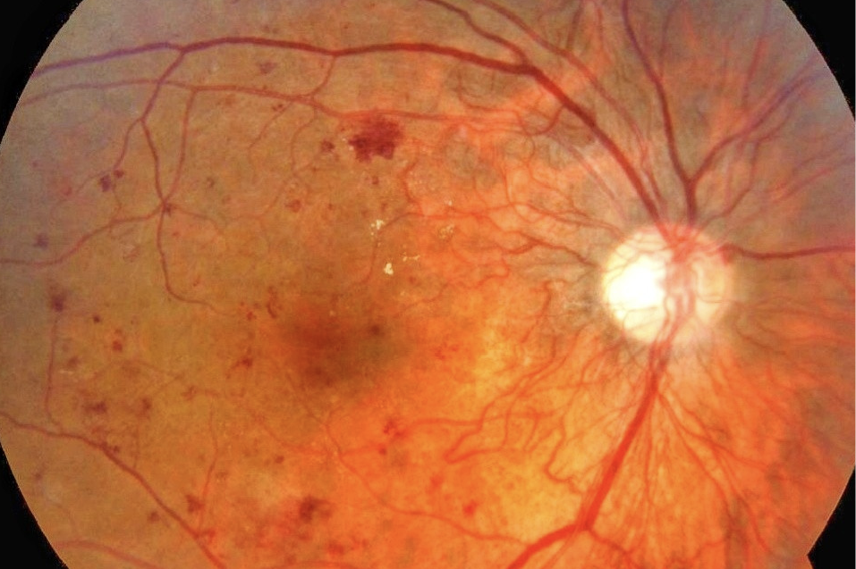
This condition often has no symptoms in its early stages. As it progresses, diabetic retinopathy may cause:

  • Blurred vision
  • Dark spots or floaters
  • Fluctuating vision
  • Impaired colour vision
  • Areas of vision loss
  • Bleeding or retinal detachment, in severe cases

Causes of Diabetic Retinopathy

The primary cause of this condition is prolonged high blood glucose levels, which cause damage to the small blood vessels in the retina. 

Other contributing risk factors include high blood pressure, high cholesterol, poor diabetic control (poor control of blood sugar levels), as well as a longer duration of having diabetes. In some cases, pregnancy may also worsen existing diabetic retinopathy.

Types of Diabetic Retinopathy

There are two main stages of this condition:
  • Non-proliferative diabetic retinopathy (NPDR): This earlier stage is marked by microaneurysms, retinal bleeding, and fluid leakage.
  • Proliferative diabetic retinopathy (PDR): This advanced stage involves abnormal blood vessel growth, which can bleed into the eye (vitreous haemorrhage), cause scarring, and increase the risk of retinal detachment.
Diabetic macular edema (DME) can occur at either stage, where fluid accumulates in the macula and affects central vision. If left untreated, DME can lead to irreversible damage of the macula and permanent vision loss.

Treatment options for Diabetic Retinopathy

Treatment depends on the severity and stage of the conditions. In the early stages, careful management of underlying health factors such as blood sugar, blood pressure, and cholesterol may slow progression.

For more advanced stages, anti-VEGF injections are commonly used to reduce swelling and prevent the growth of abnormal vessels. 

In some cases, steriod injections or implants may be recommended, particularly for diabetic macular edema.

Laser therapy (focal or pan-retinal photocoagulation) may be performed to seal leaking vessels to shrink abnormal ones.

If there is significant bleeding into the eye or traction from scar tissue, a surgical procedure called a vitrectomy may be needed to restore and preserve vision.
Cold lasers can be paired with verteporfin to close the source of the fluid leakage in each eye.

Oral medications like Eplerenone may also help control the fluid build-up.
Intravitreal anti-VEGF injections can prevent the growth of abnormal blood vessels in PCV.

PDT and verteporfin can be used to seal the abnormal vessels causing problems through leakage
Anti-VEGF injections can prevent the growth of abnormal veins in the eye, which are a common response to the occlusion, as well as reduce swelling in the retina.

PDT and verteporfin can also be used to deal with leaky blood vessels that may develop following the occlusion.

A vitrectomy can be used to remove serious haemorrhages resulting from the occlusion of the jelly-like part of the eye.

Schedule an eye screening appointment with us to assess and treat your retinal conditions

If you suspect you have any of the conditions above, book an appointment with our team of retinal specialists for an eye screening.
BOOK AN APPOINTMENT

FAQs on Retina Disease Treatments

Tests performed during eye screening examinations for retinal issues include Amsler grid, careful dilated eye examination, fluorescein & indocyanine green angiography, and optical coherence tomography (OCT).

If available, it’s best to bring any results of previous tests and records of your medical history for your appointment.

The amount of time it takes to get results from an eye screening will vary. Each case is treated on an individual bases for thoroughness.

Recovery periods vary based on your unique circumstances and the condition and treatment applicable. For example, the usual recovery period for an anti-VEGF injection for wet AMD is usually as short as several days. Meanwhile, the recovery for a vitrectomy for an RVO-related haemorrhage could take several weeks.

Each patient is unique and your doctor can assess your case to determine how many treatments you require. Most patients with wet AMD start at 3 injections every 4 weeks, but it always varies on a case-to-case basis.

RVO can be managed, especially its side effects (like vision loss). The occlusion itself cannot be treated, however.

WhatsApp Scroll to top